Ultrasound In Pregnancy
Ultrasound In Pregnancy
Introduction
A pregnancy ultrasound is an imaging test that uses sound waves to create a picture of how a baby is developing in the womb. It is also used to check the female pelvic organs during pregnancy.
First Trimester Ultrasound
(also called a level 1 ultrasound)
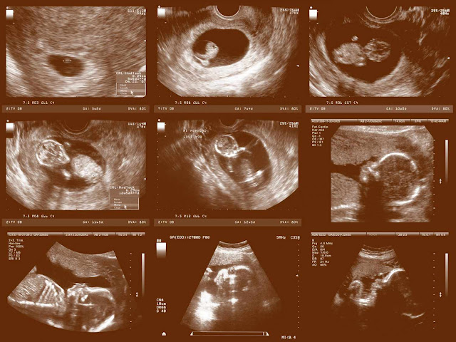
You certainly won't able to make out any adorable baby features yet (or find out whether you are boarding a boy or girl), but you are still probably pretty excited to know that earl ultra sound (at about 6-9 weeks) have become a routine part of pre-natal care, allowing eager parents a welcome first glimpse of their still tiny baby bean. The top reason why combined with LMP measurement of the embryo or fetus during early pregnancy is the most accurate way to date a pregnancy (after the first trimester ultrasound measurements of the fetus are less accurate). An easily ultrasound is also used to visualize a heartbeat, as well as to confirm that the pregnancy is taking place where it's supposed to, in the uterus (and to rule out an ectopic, or tubular pregnancy). And if you are carrying twins (or more), an early ultrasound will issue that double baby bulletin sooner.
How does a ultra sound get a picture of life inside your uterus? through sound waves emitted by a transducer wand which bounces off structures (your baby, the gestational sac and so on) to produce an image that can be viewed on a video screen. If you re getting an ultrasound before week 6 or 7, it's likely you'll have a transvaginal one.
In a transvaginal ultrasound a long narrow transducer wand (covered first with a condom like cover and sterile lubricant) is inserted into the vagina. The practitioner will gently move the wand within the vaginal canal to scan your uterus, allowing for an extra early view of your baby. After week 6-7 you'll probably be a candidate for the transabdominal version. For a trans abdominal early ultrasound, your bladder must be full (which isn't fun) so the still small uterus can be seen more easily. Gel(some practitioner warm it up first, otherwise it's chilly) is spread on your abdomen, and than the transducer wand is rubbed over it.
.png)
Both procedures can last from 5 to 30 minutes and are painless, except for the discomfort of the full bladder necessary for the first trimester trans abdominal exam and possibly the discomfort of the vaginal transducer. you'll be able to watch along with your practitioner though you'll likely need help figuring out what you're seeing) and probably take home a small print out as a souvenir.
While most Practioner will wait until at least 6 weeks to order an early ultrasound. it's possible to see a gestational sac as early as 41/2 weeks after a last menstrual period and a heartbeat as early as 5-6 weeks (thought isn't always detected.
Second Trimester Ultrasound
Anatomy Scan or (also called a level 2 ultrasound)
Get ready for the big reveal (of your baby's adorable features at least). Mom to be are routinely scheduled for an Anatomy Scan (also called a level 2 ultrasound) in the second trimester usually between 18 -20 weeks. That's because second trimester ultrasound is a great way to see how a baby is developing- and to offer reassurance that everything is going exactly the way it should be. One of the most exciting functions as far as many parents are concerned. It can give you the 411 on baby's sex, on a want to know basis, of course(that is unless you've already scored those results via an earlier chromosomal analysis). plus its fun to get a sneak peak at your baby-especially now that he or she actually looks like a baby!
Sex Determination is banned in India.
The more detailed scan will also give your practitioner additional value information about what's going on in that belly of yours. For example it can measure the size of your baby and check all the major organs. It can measure the amount of amniotic fluid to make sure there's just the right amount, and evaluate the location of your placenta. In short, the second trimester ultrasound-besides being fun to watch-will give you and your Practioner a clear picture (literally) of the overall health of your baby and your pregnancy. Eager to make some sense of what you're seeing on the screen?
Your cute beating heart will be easy to locate, but ask the sonographer to point out baby's face, hands, feet, and even some of those tiny but amazing organs, like the stomach and kidney.
 |
| 3D AND 4D Ultrasound |
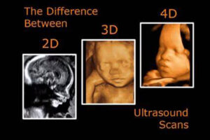
Routine second trimester ultrasound are usually done in 2D-which will provide just a flat profile of baby's feature. If a very cute one (suitable for framing or uploading)Most Practioner preserve the more detailed 3D (which takes multiple 2D images and pierce them together to form a 3D rendering that shows the whole surface, resembling a photo), and 4D scans (which shows baby moving in real time. like a video.) To more closely examine a fetus for a suspected anomaly such as cleft lip or spinal cord problems, or to monitor somethings specific that has to be seen more clearly. currently these more sophisticated (and admittedly (fun to watch) scans are officially recommended only when they're considered medically neccessary. That's because studies evaluating the safety of ultrasound technology show mixed results and as yet unclear potential risks. Thinking about springing for an upgraded in womb experience at your local prenatal portrait center ( Baby the movie) so you can get up close and personal with your little one before he or she arrives?


.jpeg)


Comments
Post a Comment